RARE IMAGING STUDIES CAN BE DONE IN OUR BEYLIKDUZU HOSPITAL.
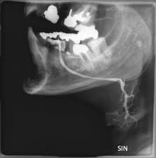
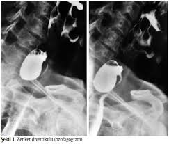
Esophagography / Faringo-Esophagography
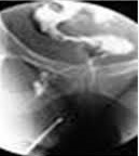
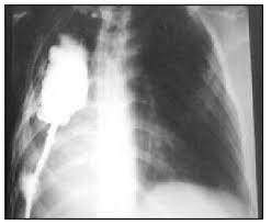
Esophagography is taken after the contrast (Color Drug) substance is given to the esophagus.
Esophagography is taken after the contrast (Color Drug) substance is given to the esophagus.
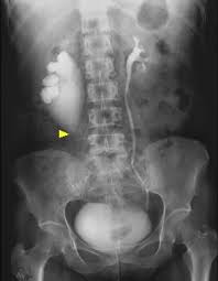
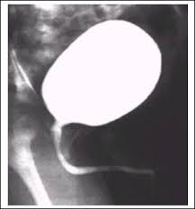
Voiding Cystouretrography / Cystogram
Voiding Cystouretrography (VSUG) is an X-ray imaging method used for the evaluation of the bladder and lower urinary system, it is the imaging of the urinary tract during voiding through the contrast agent delivered to the bladder.
Voiding Cystouretrography (VSUG) is an X-ray imaging method used for the evaluation of the bladder and lower urinary system, it is the imaging of the urinary tract during voiding through the contrast agent delivered to the bladder.
Retrograde Uretrography
Retrograde urethrography (Retrograde urethrography) is a method applied for imaging the region with x-ray by giving fluid to the urethra and determining the location and size of possible stenosis in the urethra.
Retrograde urethrography (Retrograde urethrography) is a method applied for imaging the region with x-ray by giving fluid to the urethra and determining the location and size of possible stenosis in the urethra.
Dacryocystography
It is the radiological examination of the tear sac and duct by giving contrast material.
It is the radiological examination of the tear sac and duct by giving contrast material.

FİSTULOGRAPHY
Fistula; The formation of a connection (channel) that should not normally be between the skin and intestine is called fistula.
It is the image taken by giving contrast material into the fistula.
Fistula; The formation of a connection (channel) that should not normally be between the skin and intestine is called fistula.
It is the image taken by giving contrast material into the fistula.
Pouch Chart
Loss of organ or organ cavities is the display of cavities by giving contrast agent.
Loss of organ or organ cavities is the display of cavities by giving contrast agent.
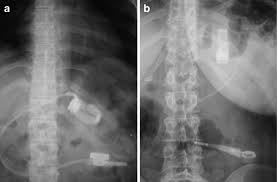
Double Contrast Column Graph
The enema is called opaque, double contrast colon chart or barium colon chart. It is the examination of large intestines by giving contrast medium and barium from breech barium.
The enema is called opaque, double contrast colon chart or barium colon chart. It is the examination of large intestines by giving contrast medium and barium from breech barium.
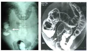
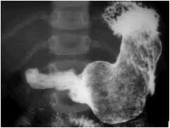
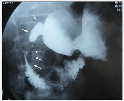
Double Contrast Stomach Graph
It is called the esophagus-stomach-duodenum (esophagus-stomach-duodenum) graph. The inner surfaces of these organs are made visible by drinking gas-containing tablets and contrast medium with barium.
It is called the esophagus-stomach-duodenum (esophagus-stomach-duodenum) graph. The inner surfaces of these organs are made visible by drinking gas-containing tablets and contrast medium with barium.
Double Contrast Small Intestine X-Ray
It is the examination of small intestines by drinking contrast material (small intestinal passage radiography) and sometimes by giving air to the patient through a tube that is swallowed (enteroclysis).
It is the examination of small intestines by drinking contrast material (small intestinal passage radiography) and sometimes by giving air to the patient through a tube that is swallowed (enteroclysis).
Sialography Graph
It is a radiography obtained after injection of a radiopaque contrast agent through the canal of the salivary gland system.
It is a radiography obtained after injection of a radiopaque contrast agent through the canal of the salivary gland system.